Anatomy Of Back Of Neck : Human Muscles of the Neck Poster - Clinical Charts and ... / The hard white exterior covering of the tooth is the enamel.
byAdmin-
0
Anatomy Of Back Of Neck : Human Muscles of the Neck Poster - Clinical Charts and ... / The hard white exterior covering of the tooth is the enamel.. Some important structures contained in or passing through the neck include the seven cervical vertebrae and enclosed spinal cord, the jugular veins and carotid arteries, part of the esophagus, the larynx Below the neck, holding the tooth into the bone, is the root of the tooth. It is designed to be incredibly strong, protecting the highly sensitive nerve roots, yet highly flexible, providing for mobility on many different planes. The cervical spine supports the weight and movement of your head and protects the nerves exiting your brain. Spinal anatomy is a remarkable combination of strong bones, flexible ligaments and tendons, large muscles and highly sensitive nerves.
Cervical spinal stenosis is a narrowing of the middle portion of the spinal canal and can lead to compression of the spinal cord. Apr 12, 2018 · the image below to shows all the major back muscles (as well as some neck muscles): The neck is resilient enough to sustain a five kilogram weight 24/7, yet sufficiently mobile to move it in several directions. The splenius muscles originate at the midline and run laterally and superiorly to their insertions. Neck, in land vertebrates, the portion of the body joining the head to the shoulders and chest.
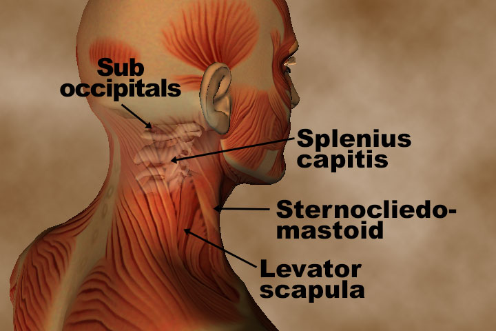
Anatomy and Pathology for bodyworkers - Real Bodywork from www.realbodywork.com The neck is resilient enough to sustain a five kilogram weight 24/7, yet sufficiently mobile to move it in several directions. Still, many individuals pay far too little attention to them. From the sides and the back of the neck, the splenius capitis inserts onto the head region, and the splenius. It is designed to be incredibly strong, protecting the highly sensitive nerve roots, yet highly flexible, providing for mobility on many different planes. The back muscles stabilize and move the vertebral column, and are grouped according to the lengths and direction of the fascicles. The rounded upper projections of the back teeth are cusps. Below the neck, holding the tooth into the bone, is the root of the tooth. Jul 16, 2019 · the neck muscles, including the sternocleidomastoid and the trapezius, are responsible for the gross motor movement in the muscular system of the head and neck.
Apr 12, 2018 · the image below to shows all the major back muscles (as well as some neck muscles):
This conditions is called a cervical radiculopathy. From the sides and the back of the neck, the splenius capitis inserts onto the head region, and the splenius. Nerve compression may cause pain that radiates (travels) from the neck into the upper back and arm(s). Apr 12, 2018 · the image below to shows all the major back muscles (as well as some neck muscles): The rounded upper projections of the back teeth are cusps. Cervical spinal stenosis is a narrowing of the middle portion of the spinal canal and can lead to compression of the spinal cord. Jul 24, 2021 · the content of the neck is grouped into 4 neck spaces, called the compartments. Still, many individuals pay far too little attention to them. Below the neck, holding the tooth into the bone, is the root of the tooth. The hard white exterior covering of the tooth is the enamel. Jul 16, 2019 · the neck muscles, including the sternocleidomastoid and the trapezius, are responsible for the gross motor movement in the muscular system of the head and neck. The back anatomy includes some of the most massive and functionally important muscles in the human body. Neck, in land vertebrates, the portion of the body joining the head to the shoulders and chest.
Jul 24, 2021 · the content of the neck is grouped into 4 neck spaces, called the compartments. It is designed to be incredibly strong, protecting the highly sensitive nerve roots, yet highly flexible, providing for mobility on many different planes. From the sides and the back of the neck, the splenius capitis inserts onto the head region, and the splenius. The cervical spine supports the weight and movement of your head and protects the nerves exiting your brain. Cervical spinal stenosis is a narrowing of the middle portion of the spinal canal and can lead to compression of the spinal cord.
Muscles of the Head and Neck Poster 24 x 36 from www.acupunctureproducts.com Nerve compression may cause pain that radiates (travels) from the neck into the upper back and arm(s). Contains glands ( thyroid , parathyroid, and thymus ), the larynx , pharynx and trachea. As the tooth tapers below the gumline, the neck is formed. The cervical spine supports the weight and movement of your head and protects the nerves exiting your brain. Cervical spinal stenosis is a narrowing of the middle portion of the spinal canal and can lead to compression of the spinal cord. The splenius muscles originate at the midline and run laterally and superiorly to their insertions. The rounded upper projections of the back teeth are cusps. Some important structures contained in or passing through the neck include the seven cervical vertebrae and enclosed spinal cord, the jugular veins and carotid arteries, part of the esophagus, the larynx
Contains glands ( thyroid , parathyroid, and thymus ), the larynx , pharynx and trachea.
The cervical spine supports the weight and movement of your head and protects the nerves exiting your brain. Cervical spinal stenosis is a narrowing of the middle portion of the spinal canal and can lead to compression of the spinal cord. Nerve compression may cause pain that radiates (travels) from the neck into the upper back and arm(s). Apr 12, 2018 · the image below to shows all the major back muscles (as well as some neck muscles): Still, many individuals pay far too little attention to them. Jul 24, 2021 · the content of the neck is grouped into 4 neck spaces, called the compartments. Below the neck, holding the tooth into the bone, is the root of the tooth. As the tooth tapers below the gumline, the neck is formed. May 31, 2021 · head and neck (anterior view) the head and neck are two examples of the perfect anatomical marriage between form and function, mixed with a dash of complexity. This conditions is called a cervical radiculopathy. The back anatomy includes some of the most massive and functionally important muscles in the human body. The back muscles stabilize and move the vertebral column, and are grouped according to the lengths and direction of the fascicles. They move the head in every direction, pulling the skull and jaw towards the shoulders, spine, and scapula.
The back muscles stabilize and move the vertebral column, and are grouped according to the lengths and direction of the fascicles. Contains glands ( thyroid , parathyroid, and thymus ), the larynx , pharynx and trachea. The hard white exterior covering of the tooth is the enamel. Cervical spinal stenosis is a narrowing of the middle portion of the spinal canal and can lead to compression of the spinal cord. Nerve compression may cause pain that radiates (travels) from the neck into the upper back and arm(s).
Acute Wry Neck (Torticollis) - Thermoskin - Supports and ... from www.thermoskin.com Below the neck, holding the tooth into the bone, is the root of the tooth. The hard white exterior covering of the tooth is the enamel. It is designed to be incredibly strong, protecting the highly sensitive nerve roots, yet highly flexible, providing for mobility on many different planes. They move the head in every direction, pulling the skull and jaw towards the shoulders, spine, and scapula. Neck, in land vertebrates, the portion of the body joining the head to the shoulders and chest. Apr 12, 2018 · the image below to shows all the major back muscles (as well as some neck muscles): May 31, 2021 · head and neck (anterior view) the head and neck are two examples of the perfect anatomical marriage between form and function, mixed with a dash of complexity. The splenius muscles originate at the midline and run laterally and superiorly to their insertions.
The hard white exterior covering of the tooth is the enamel.
The cervical spine supports the weight and movement of your head and protects the nerves exiting your brain. The splenius muscles originate at the midline and run laterally and superiorly to their insertions. It is designed to be incredibly strong, protecting the highly sensitive nerve roots, yet highly flexible, providing for mobility on many different planes. Spinal anatomy is a remarkable combination of strong bones, flexible ligaments and tendons, large muscles and highly sensitive nerves. The back muscles stabilize and move the vertebral column, and are grouped according to the lengths and direction of the fascicles. They move the head in every direction, pulling the skull and jaw towards the shoulders, spine, and scapula. The inner portions of the tooth consist of the dentin, a bonelike tissue, and the pulp. Jul 16, 2019 · the neck muscles, including the sternocleidomastoid and the trapezius, are responsible for the gross motor movement in the muscular system of the head and neck. Contains glands ( thyroid , parathyroid, and thymus ), the larynx , pharynx and trachea. As the tooth tapers below the gumline, the neck is formed. Still, many individuals pay far too little attention to them. Apr 12, 2018 · the image below to shows all the major back muscles (as well as some neck muscles): Nerve compression may cause pain that radiates (travels) from the neck into the upper back and arm(s).